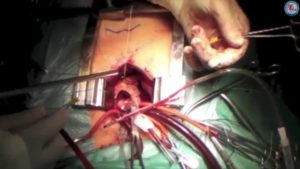
COMPLEX CORONARY BYPASS SURGERY WITH AORTIC VALVE REPLACEMENT
- drpavankumar
- 0
- on Aug 08, 2017

Case Mrs. J. F. 67 year old female had long standing Aortic stenosis since last 10 years , managed medically. This gradually progressed to severe category since last 6 months with Echo showing Aortic Valve area of 0.8 cm2 and Aortic Valve gradient of 110 MMHg with heavily calcified Aortic valve . Coronary Angiography showed severe triple vessel, vessel involving 90% block, LAD – 90% blocked OM2 & OM2 with 80% RCA ostial stenosis. Left ventricular ejection fraction on Echo was 45% with moderate L.V. hypertrophy. CT scan of Ascending aortic showed extension of heavy calcification to coronary ostia. Aortic valve replacement in this scenario become very complicated in operating strategy – due to 1) small Aortic root & annulus 2) Heavy Aortic calcification 3) Triple vessel coronary
Artery disease 4) compromised L.V. function.
With small aortic root with Aortic annulus size of 1.8cm2, It was decided to do this case on cardiopulmonary bypass & cardioplegic arrest through Retrograde cardioplegia & myocardial protection protocol by graft perfusion by doing Triple vessel CABG ( LIMA – LAD & saphenous vein grafts to OM1 & OM2 & Saphenous vein to RCA ) & Vein graft cardioplegic Myocardial protection during AVR .
As per protocol patient put on CPB with standard Aortic &caval cannulation & Retrograde cardioplegia (RCP) cannula insertion. After aortic clamping cardiac arrest achieved by RCP followed by coronary revascularisation. standard Aortatomy was made to visualise Aortic valve. As expected it was heavy calcified valve with calcification extending deep in annulus with hardly visible coronary ostia. Carefully decalcification was carried out with excision calcified valve. Aortic annular size measured to be 18mm only. As no Tissue valve of < 19mm is available, 17mm mechanical bileaflet prosthetic valve was implanted successfully , As patients body weight was 50 kg only, it was thought to be adequate valve size for this body weight. Aortatomy closed. Proximal anastomosis carried out on Aorta. C.P.B. terminated after carefully deairing with intra op TEE guidance.
Operation completed with good hemodynamic state. Patient weaned off ventilator next day in ICU & discharged on 7th day post operative day.
Discussion – Successful AVR in small Aortic root & Aortic annulus & coronary artery disease depends on specific protocols for these complexities. Good Myocardial protection, coronary perfusion & global revascularisation careful decalcification of aortic valve for adequate sized aortic prosthetic valve implantation ( without need of aortic root widening) gives excellent out come . Experience to avoid complications & avoidance of overzealous practices are essential.


