
PAPVC + SINUS VENOSUS DEFECT HEART SURGERY.
- drpavankumar
- 0
- on Sep 07, 2022
Mr. IK 42 years male complained about breathlessness on exertion since last 2 years . On
investigations chest X’ray chest showed moderate enlarged heart & routine Echo cardiogragrapy
Showed high pulmonary artery pressure artery pressure & right ventricular overload, but failed to
show any congenital hole in the heart. Later transesophegeal Echo showed up sinus venosus type
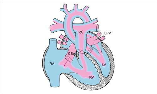
Atrial Septal Defect (ASD). CT scan pulmonary angiography showed that Right superior & middle
pulmonary veins draining into right atrium via Supeior vena cava( SVC) making a diagnosis of right
partial Anomalous Pulmonary venous Connection ( PAPVC).
Cardiac catheterisation on patient showed predominant left to right shunt across sinus venosus ASD
with severely high Pulmonary Artery pressure .
Patient was taken up for Open Heart Surgery repair on 8 March’2019 at BSES hospital by Dr. Pavan
Kumar. During his surgery findings showed that he had 4.5 x 4.5 cm2 large sinus venous ASD with
patent foramen Ovale ( PFD ). PAPVC drained into SVC. 6cm Large DACRON Patch was then used
to Re-route PAPVC drain into left Atrium with closure of Sinus Venosus ASD which included of
PFO repair. Surgery went off very well & patient had uneventful recovery.
Discussion – Sinus Venosus ASD are less common variety of Secundum ASD disease less than 5%
patient have sinus venosaus ASD of all secundum variety. In about 50% such cases Right PAPVC is
found.
These are difficult to diagnose by normal Echo . CT scan chest with pulmonary angiogram is usually
diagnostic of PAPVC. Surgical correction is only treatment of choice with assured success.